Each year Rein in Sarcoma’s Jan Maudlin Sarcoma Scholars complete a project involving sarcoma cancer education and awareness. In 2015, some of the scholars conducted assessments of Minnesota primary care practitioners at two educational conferences. Their work, summarized by Dr. Croix Fossum, a scholar from Mayo Clinic and now at the Keck School of Medicine at USC, Los Angeles, CA, was published in the April 2020 Mayo Clinic Proceedings: Innovations, Quality & Outcomes.
We want to thank Dr. Scott Okuno for assisting with the publication. He is a Rein in Sarcoma Board Member, Research Committee member, and oversees the Jan Maudlin Sarcoma Scholars at the Mayo Clinic Alix School of Medicine.
The assessment concluded that “Minnesota PCPs have seen very few cases of sarcoma and report low familiarity with sarcoma guidelines. When challenged with case presentations, PCPs made decisions inconsistent with established guidelines. This study supports ongoing efforts to increase sarcoma awareness.”
Propelled in part by this study, the RIS Red Flags of Sarcoma Education Committee significantly expanded their education efforts. They work with the RIS Medical Advisory Board to educate primary care practitioners (PCPs), school nurses and athletic trainers, at several conferences each year. The committee also oversees the Hallie Anne Brown Initiative, which is working to develop and evaluate the effectiveness of best practice alerts in improving sarcoma diagnosis accuracy and timeliness.
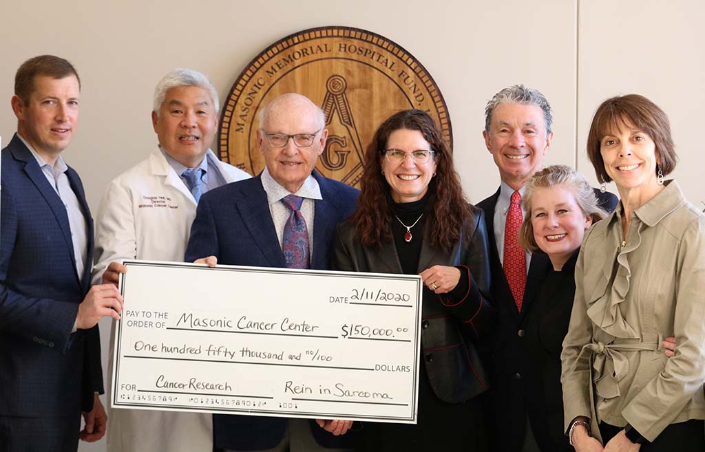
Rein in Sarcoma proudly announces a $150,000 research grant approved by the Rein in Sarcoma Board on January 27, 2020. This brings the cumulative research funding by Rein in Sarcoma to $2 million since the organization’s founding in 2001. This grant will fund the following three exciting University of Minnesota Masonic Cancer Center projects:
“eBAT as a Modulator of the Myeloid Immune Checkpoint in Cancer.” | $50,000
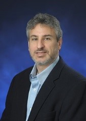
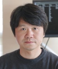
Dr. Jaime Modiano
Principle Investigator: Jaime Modiano, VMD, PhD
Co-Investigator: Jong Kim, VMD, PhD
Summary: eBAT (EGF bispecific angiotoxin) is a drug with excellent safety profile that has shown efficacy against sarcomas in vitro, in small laboratory animal models, and in dogs with spontaneous vascular sarcomas. Our goal is to determine eBAT’s mechanisms of action to rationally expand its use to treat human sarcomas where it will provide comparable benefits and address a critical unmet need. The hypothesis is that eBAT eliminates immunosuppressive myeloid cells in the tumor environment, promoting enhanced anti-tumor immunity. We will test the hypothesis through one aim, to determine the effect of immunosuppressive myeloid cell depletion or persistence in the therapeutic efficacy of eBAT against sarcomas. The experimental model will consist of syngeneic mouse fibrosarcoma, where we have deleted the urokinase receptor (uPAR) using genome editing, uPAR-knockout mice, and a novel methodology to generate bone marrow chimeras in newborn mice which will promote full donor chimerism of myeloid cells, lymphoid cells and tissue macrophages. eBAT, and its mouse specific homolog, meBAT, will be tested in this model to examine their ability to induce tumor responses and immune infiltration in animals with wild type and uPAR-deficient tumors implanted in wild type mice and in chimeric mice with uPAR-knockout myeloid cells and macrophages. We predict that eBAT will have modest effects to reduce tumor growth in this model, due the low affinity binding to mouse UPAR. On the other hand, we expect meBAT will reduce growth of uPAR+ tumors in all recipients, but will only reduce growth of uPAR-KO tumors in mice with uPAR+ bone marrow. We anticipate tumor reduction will be associated with fewer myeloid-derived suppressor cells and macrophages in the tumors, as well as increased numbers of infiltrating T cells and NK cells. This experiment will allow us to define appropriate conditions for subsequent experiments to study the immune response in greater depth and to test the effect of eBAT in combination with drugs that block the PD-1/PD-L1 T-cell exhaustion checkpoint.
“Using Propranolol to Generate an Anti-Tumor Microenvironment.” | $50,000
Summary: Macrophages can be programmed within the tumor microenvironment toward anti-tumor or pro-tumor responses. Tumor cells can promote the pro-tumor functions of macrophages by scavenging cholesterol from the membranes of macrophages. In contrast, cholesterol accumulation in macrophages promotes an anti-tumor phenotype. We recently found that propranolol, a drug commonly used to treat heart disease, disrupts the ability of tumor cells to scavenge extracellular substrates such as cholesterol. Based these findings and our preliminary data, we propose that propranolol inhibits tumor cells from scavenging cholesterol from tumor associated macrophages, programming macrophages toward an anti-tumor phenotype and harnessing their ability to promote adaptive, anti-tumor immune responses. Studies are proposed to determine if propranolol prevents sarcoma cells from scavenging cholesterol from macrophages, and whether propranolol promotes a shift toward an anti-tumor microenvironment. Using a combination of in vitroand in vivoapproaches, these studies will provide novel information regarding the ability of propranolol to remodel the tumor microenvironment, resulting in decreased immune suppression and enhanced anti-tumor responses. Successful completion of the proposed work investigating the positive impact of propranolol on reprogramming of the tumor microenvironment will accelerate our progress in the identification of synergistic drug combinations and the inclusion of tumor immunotherapies.
“Computationally Deciphering the Paths of Genomic Catastrophe in Osteosarcoma.”| $50,000
Summary: Osteosarcoma is characterized by massive genomic catastrophes. Timing and relative ordering between the genomic catastrophic constraints should allow both earlier diagnosis and better prediction of tumor progression. However, such evolutionary trajectories remain elusive due to the lack of advanced computational methods that robustly deduce the paths of somatic changes from next-generation sequencing (NGS) data. Drawing on the full spectrum of somatic alterations detectable from NGS data, we seek to fill a lacuna in knowledge on cancer evolution through innovating reconstructive computational algorithms to decipher the time ordering of genomic catastrophe in osteosarcoma. Our algorithms will enable the inference of the relative ordering of loss of heterozygosity, whole genome doublings, structural changes and other localized catastrophes in osteosarcoma using public NGS data. Backtracking the chaos to discover the initiating genomic event may reveal novel drivers of this devastating disease and strategies to therapeutically intervene. The mode of evolutionary trajectories of catastrophes is a key genomic feature allowing better patient stratification in terms of tumor evolvability. The computational framework established here, whereas inspired by the complexity of the OS genome, will have an immeasurable impact on tracking the dynamics of other sarcoma subtypes showing genome instability, particularly given the urgent need to account for copy number alterations in studies of tumor evolution.
Rein in Sarcoma has awarded $45,000 in new sarcoma research grants to Children’s Minnesota and the Mayo Clinic.
Children’s Minnesota
“Expanding Opportunities For Early Detection of Sarcoma.” | $20,000
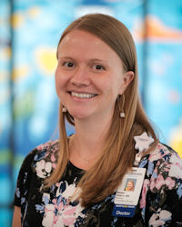
Dr. Megan Hilgers
Dr Kris Ann Schultz
Co-Principal Investigators: Megan Hilgers, MD, pediatric oncologist and Kris Ann P. Schultz, MD, pediatric oncologist.
Lay Summary: Liquid biopsies are a promising technique to facilitate tumor monitoring and, in certain clinical circumstances, early detection. In this application, we propose to build on our existing ctDNA work in DICER1-related tumors and expand ctDNA research at Children’s Minnesota to include non-DICER1 related sarcomas including Ewing’s sarcoma, osteosarcoma and the variety of aggressive tumors including sarcomas which are seen at high frequency in children and adults with Li Fraumeni syndrome.
Funding from Rein in Sarcoma will: 1) Facilitate our participation as founding members of the Early Detection of Inherited and SecondarY Neoplasms (EDISYN) Consortium; and 2) Allow us to open a trial to assess the impact of ctDNA levels on survival in children with Ewings Sarcoma and osteosarcoma. Both of these projects leverage the clinical strengths of Children’s Minnesota and our existing DICER1related ctDNA research with existing laboratory expertise outside of Minnesota to more expeditiously bring ctDNA-related studies to children with sarcoma in Minnesota and worldwide.
Mayo Clinic
“Integrated Proteomic and Transcriptomic Profiling of Rhabdomyosarcoma Reveals Target Antigens for Immune-Based Therapies.” | $25,000
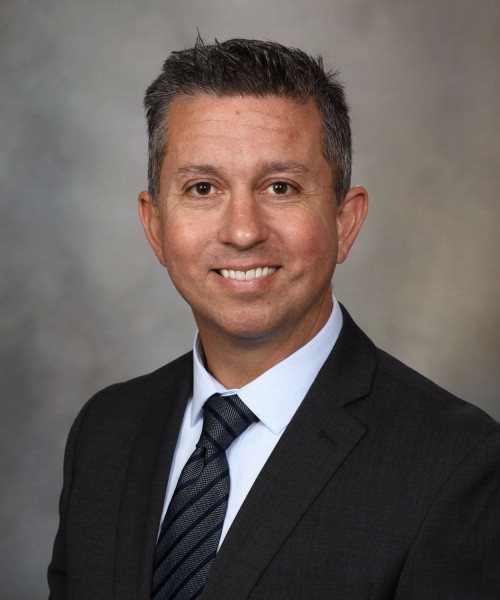
Dr. Patricio Gargollo
Principal Investigator: Patricio Gargollo, MD, Associate Professor in Urology
Co-Principal Investigators: Dr. Carola Arndt, MD, Professor of Pediatrics, Dr. Candace Granberg, MD, Assistant Professor of Urology, Dr. Haidong Dong, MD, PhD, Professor of Immunology, Dr. Fabrice Lucien-Matteoni, PhD, Senior Research Fellow in Urology
Lay Summary: Rhabdomyosarcoma (RMS) is the most common soft tissue tumor in children, with nearly 20% of children presenting with locally aggressive and/or metastatic disease. A fundamental problem with this disease is the lack of effective and tolerable therapeutic regimens. Current protocols including surgery, radiotherapy and chemotherapy are extremely toxic and may lead to multiple deleterious long-term effects. Moreover, a significant percentage of patients tends to relapse and for those patients, life-expectancy is less than 5 years. Our group is dedicated to uncover molecules at surface of tumor cells aberrantly expressed in RMS. The biggest benefit of our approach is that cell-surface proteins are easily accessible for pharmacological intervention as cell surface is an effective barrier for the entry of therapeutic drugs. Our first objective is to analyze the repertoire of cell-surface molecules in RMS tumors and compare with normal muscle to identify potential targetable vulnerabilities. We will examine the functional impact of the expression of RMS-specific molecules on tumor growth using patient-derived RMS cells.
This proposal will lay the foundation for our next short-term goal which will be to design new therapeutic strategies to target RMS-specific molecules and evaluate their clinical potential for the treatment of pediatric RMS. Ultimately, our study will expand the therapeutic landscape of children diagnosed with RMS by improving patient response to treatment, increasing their survival rate and reducing treatment-related toxicities. Finally, our successful approach will be suitable to overcome similar challenges in other types of sarcoma very aggressive in children such as osteosarcoma and Ewing sarcoma.
These grants are made in addition to research grants to the University of Minnesota made in January of this year.
Beau Webber, PhD – Assistant Professor, Department of Pediatrics, University of Minnesota
Beau Webber, a 2018 RIS research grant recipient, is an Assistant Professor, Department of Pediatrics at the University of Minnesota, as well as Faculty Member of the Division of Pediatric Hematology and Oncology and Stem Cell Institute. While getting his PhD in stem-cell biology and genetic engineering at the University of Minnesota, Webber used genetic tools to help correct mutations that cause disease. He worked primarily with cells called Induced Pluripotent Stem Cells (iPSCs). Basically, iPSCs are adult cells that have been genetically reprogrammed into embryonic stem cells, helping eliminate the ethical concern with using real embryonic cells.
While working with a colleague, Webber was introduced to cancer genetics, which included understanding which genes go wrong in a cell that leads to cancer. Webber’s colleagues created mouse models to do forward genetic screens by using a transposon system, or something called a jumping gene that would jump around and land in genes, causing the cell to become cancerous. Working backwards, Webber’s team studied these genes in the mice to determine which genes became broken, causing cancer. Then his team would identify cells that could be targeted with specific therapies. However, mice and humans are very different from one another, limiting the usefulness of mice studies. Another way to study cancer is to put tissue samples from human tumors in a culture dish and see which cells stick and grow with the right conditions. This would show which lines were cancer. While this works, it’s a poor representation of what tumors look like considering tumors are three-dimensional and complex. So, Webber’s goal became how can we make these tumors more realistic and “better predictors” to help with treatments. By using what he was familiar with, Webber hoped to make models of cancer using iPSCs cells. He wanted to combine what his colleagues were doing in the mice with the iPSCs cells, to create a controlled way to make the iPSCs cells into cancer. Through this process, Webber wants to understand all the different pieces that need to be broken in order to make the iPSCs cells into cancer.
By using a ground up method, Webber starts with a normal iPSCs cell and then breaks it piece by piece until he gets something that resembles cancer. There’s a lot of potential with this method in that they can observe the cell becoming cancer in a dish. The next step involves genetic engineering. Currently there are a lot of techniques to help make 3D models resembling cubes or a circular shape. These are known as organoids. These organoids can take on all the complex properties associated with organs. By using genetic engineering to help create 3D shapes and by using iPSCs cells, Webber is hoping to create mini 3D tumors that can be easily monitored. By creating these through a controlled process, Webber’s’ team will know everything that “went wrong” (each step required to create the cancerous tumor).
Webber is currently working on two main projects involving Osteosarcoma and Ewing Sarcoma. While the two cancers differ from one another, they start off virtually the same—a bone progenitor cell. First, Webber and his team direct the iPSCs cells to become mesenchymal progenitor cells, which then can become cartilage and bone. Webber created his own serum to help ensure that every step was as controlled as possible. After, “getting those systems to work really well” the next steps consisted of genetic engineering. There are well known proteins called tumor suppressors – think of these proteins as breaks. Webber and his team use CRISPR technology, a system that was taken from bacteria that help fight off viruses. CRISPR is used as a way to cut DNA wherever needed. Webber and his team can cut these tumor suppressors in the iPSCs cells. Then they add oncogenes, genes that have the potential to turn tumorous. By cutting the breaks, the tumor suppressors, and by applying the gas, adding the oncogenes, Webber’s team is modifying the bone IPS cells to try to replicate Osteosarcoma. They are getting pretty close. In the mice screening, Webber’s colleagues believe they found genes that could be linked to Osteosarcoma metastasis. Once the model is created, Webber can put those genes in the 3D Osteosarcoma model and see how it influences the cell.
With Ewing Sarcoma it is a bit different. A unique characteristic associated with Ewing Sarcoma is nearly every diagnosed patient is of European dissent. A common mutation that occurs in Ewing Sarcoma is called the EWS-FLI-1 fusion translocation. This is present in 90% of Ewing cases. There is only one driving fusion protein. Webber describes this as, “[The protein] scrambles the computer programming of a cell. It jumps around and turns on genes that shouldn’t be on and shuts genes off that shouldn’t be off.” By using iPSCs cells, Webber’s team used CRISPR to cut at EWS and at FLI to break them and then get them to come together in the cell, creating that EWS-FLI-1 translocation, leading to Ewing Sarcoma. The EWS-FLI-1 protein when injected into most cells kills them. Webber and his team are working on finding the perfect balance between turning the protein on at a high level while keeping the cell alive. They are aiming to hit that fine line in a specific cell type in a given situation where the cell can respond to the protein being turned on. For all this to occur the perfect condition must be present.
Currently with Ewing Sarcoma, Webber’s team is working on inserting these EWS-FLI-1 translocations into the different cell lines to see which lines stick and start forming Ewing Sarcoma in the bone cells made from iPSCs cells. They are looking to answer, “What is the exact dose that is required to get these cells to transform?” Then by utilizing the system, they can determine the origin of Ewing Sarcoma. Using targeted therapies, Webber and his team can then see how the tumor responds to the given therapy.
Using the Rein in Sarcoma research grant, Webber went to the WiCell lab in Madison, Wisconsin. There, they pulled out 10 iPSCs lines ranging from 100% European to 100% African ancestry and many degrees in between. Webber and his team are hoping to eventually convert those lines to Ewing Sarcoma. What most likely will happen is the European cell lines will turn into Ewing Sarcoma and those of African ancestry won’t. This may or may not occur, but the goal is to monitor what happens when the EWS-FLI 1 protein is turned on in the differing ancestry lines. Webber’s team believe that a comparison of the two ancestry lines will help point to the important genes that potentially cause Ewing Sarcoma Ultimately, using his prior knowledge of iPSCs cells, Webber is hoping to learn more about the formation of sarcoma cancers to determine the most beneficial treatments. By striving to create artificial sarcoma tumors, more research can be done, since patient samples are no longer the only way to obtain tumor samples.
Currently, Webber and his team are satisfied with the progress being made towards the final goal of changing these iPSCs cells into tumors. While they are not completely there, Webber and his team are well on the way to providing new tools and understanding towards the fight against sarcoma cancer.
Miranda Mead is a Ewing Sarcoma survivor and Journalism student at the University of St. Thomas. As an RIS volunteer, Miranda is interviewing RIS research grant awardees, such as Dr. Webber, to bring that work to life for the Rein in Sarcoma community. Read Miranda’s sarcoma story
Funding sarcoma research is one prong of Rein in Sarcoma’s three-pronged mission. With the great support of donors including a significant gift through the Hallie Anne Brown Named Fund, physicians, researchers, and board members RIS expanded research funding in 2017. This expansion not only grew our commitment to the University of Minnesota, but provided research grants to Children’s Hospital, Minneapolis and the Mayo Clinic, Rochester.
Dr. Scott Okuno, Dr. Brenda Weigel and Dr. Larry Seymour
At that time the Rein in Sarcoma Research Committee was formed to oversee RIS research grant awards. To provide a forum for researchers to present findings to RIS stakeholders and further the collaboration among RIS sponsored researchers, the committee initiated the first RIS Researcher Symposium, held at the UMN Cancer and Cardiovascular Research Building on April 27. Co-hosted by committee chair, Dr. Larry Seymour, Peter Wyckoff, and Dr. David Largaespada, the symposium was incredibly successful and we look forward to another in 2020!
After breakfast with the committee and RIS Board members, the 2017 research grant recipients presented their work to each other and Research Committee members. At the end of the morning, Dr. David Largaespada led board and Research Committee members on an captivating tour of his sarcoma research lab. We met and heard from four student researchers about their work in finding the mechanisms and potential cures to sarcoma cancers.
Thanks to Dr. Largaespada and his colleagues for hosting the symposium and providing the tour, with special thanks to Emerson Fuller for her detailed coordination assistance. We appreciate the presenting researchers’ preparation and willingness to participate. Thanks also to Bill Donovan for photographing the morning.
Presenting Researchers: L. Chinsoo Cho, MD, Erin Dickerson, PhD, Avudaiappan Maran, PhD, Kris Ann Schultz, MD, Jessica Lawrence, DVM
UMN Sarcoma Lab Researchers: Minu Bhunia, Bryant Keller, Emily Pope, Julia Nikrad
RIS Research Committee: Dr Larry Seymour, Brenda Weigel, MD, David Largaespada, PhD, Kris Ann Schultz, MD, Scott Okuno, MD, Keith Skubitz, MD, Peter Wyckoff, Janelle Calhoun
On May 2019, Mayo Clinic oncologist and RIS Medical Advisory Board Member, Dr. Steven Robinson, hosted a meeting for Mayo colleagues to learn about Rein in Sarcoma’s initiatives. Over lunch provided by the Red Flags Committee, Mayo Radiation Oncologist and RIS Medical Advisory Board Member Safia Ahmed, presented the new Rein in Sarcoma Patient Starter notebook. Dr. Ahmed and other Medical Advisory Board members provided new information and helped the Red Flags Committee bring the notebook up to date.
RIS Maudlin Sarcoma Scholars from Mayo, Taylor Weikittel and Mylan Blomquist, told about the many activities they have organized for Mayo Medical School Students to raise sarcoma awareness. They also summarized the survey they and the University of Minnesota scholars completed – asking former scholars about their experiences after their sarcoma scholar year. Kara Dolney, Mayo patient and RIS Patient and Family Support Committee member, spoke about her sarcoma experience and the RIS support available to Mayo sarcoma patients.
Dr. Steven Robinson
Pete Wyckoff and Dr. Robinson summarized the RIS research grants to Mayo and then led a discussion about further collaboration. Mayo Medical Staff attendees were very attentive and asked many questions. Molecular Pathologist Kevin Halling wrote the next day to say how much he enjoyed the meeting:
“As a pathologist I don’t often get to see the direct patient care side of things and so I found yesterday’s meeting very enlightening. Again, thanks for all your work.”
Kevin Halling, Molecular Pathologist, Mayo Clinic
Doctors and staff from departments that had not previously handed out Rein in Sarcoma Patient Notebooks eagerly took notebooks we had brought to start giving to their patients. Rein in Sarcoma and Mayo Clinic enjoy a great collaboration that encompasses Rein in Sarcoma’s three-pronged mission: sarcoma education, patient support, and funding sarcoma research. It was a great day of sharing ideas for all of us.